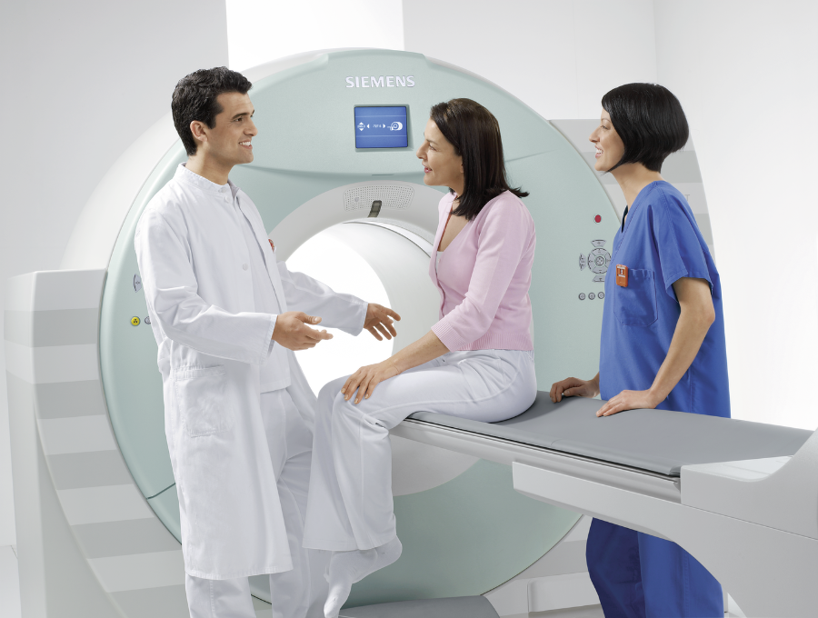
Bone Imaging
A PET/CT scan with sodium fluoride F 18 injection (18F NaF) is a nuclear imaging test that scans the entire skeletal system and produces high-resolution images of the bones. These images are used to detect areas of abnormal bone growth associated with tumors.
A bone scan is an important tool for detecting cancer that has metastasized or spread to the bone from a tumor that started in a different organ, such as the breast or prostate.
Some of the most prevalent cancers in the United States are commonly associated with metastatic bone disease. This is of particular clinical importance in breast and prostate cancers because of the prevalence of these diseases. At postmortem examination, 70% of breast and prostate cancer patients had evidence of metastatic bone disease.1 However, bone metastases are not restricted to only these two cancers. They may complicate a wide range of other malignancies, resulting in considerable morbidity and complex demands on health care resources. Carcinomas of the thyroid, kidney, and lung also commonly give rise to bone metastases, with an incidence at postmortem examination of 30% to 40%.1
18F NaF bone imaging provides the physician with physiologic information of the bone. Live adult bone is not a rigid inorganic framework. At millions of microscopic sites throughout the skeleton, and especially at areas of disease, bone is constantly being broken down and then remade in a cellular process termed remodeling. When the PET scan shows areas of increased uptake of 18F NaF in the skeleton, it reflects sites of increased blood flow and bone remodeling. This information can be used by physicians to diagnose bone disease, detect bone injury or determine the extent of metastatic disease.
A bone PET/CT scan’s high-resolution images and its ability to scan the entire skeleton make it very helpful in detecting areas of abnormal bone growth associated with tumors. The test poses no greater risk than do conventional x-ray procedures, as the radiopharmaceutical used produces very little radiation exposure.
Effective February 2011, the Centers for Medicare and Medicaid (CMS) began covering 18F NaF PET scans as part of the National Oncologic PET Registry (NOPR).
References:
- Galasko C. The anatomy and pathways of skeletal metastases. In: Weiss L, Gilbert A, editors. Bone metastases. Boston: GK Hall; 1981. p. 49 – 63.